Brain Age vs. Chronological Age: The Science of Cognitive Longevity and Mental Aging
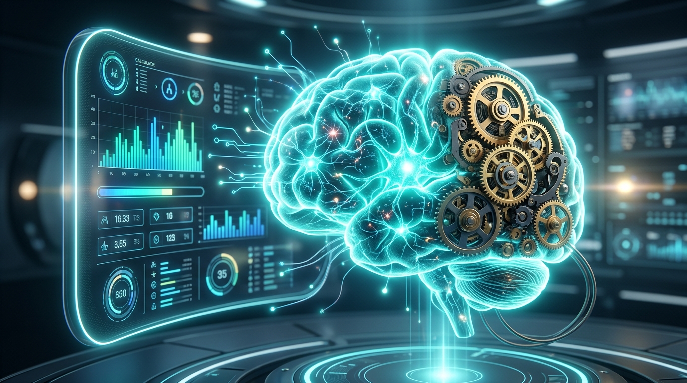
Brain Age vs. Chronological Age: The Science of Cognitive Longevity and Mental Aging
For centuries, humanity has measured aging through a single, unyielding metric: the relentless tick of the astronomical calendar. We celebrate birthdays, calculate our retirement milestones, and judge our biological standing based on the number of times the Earth has completed its orbit around the Sun. This number is our Chronological Age.
However, modern cognitive neuroscience is revealing a far more complex, profound, and exciting reality: our biological tissues do not age at a uniform rate. Nowhere is this divergence more striking—or of higher consequence—than in the human brain. While two individuals may both celebrate their 60th birthday, sharing the exact same chronological age, one may possess the vibrant, highly connected, and resilient brain of a 40-year-old, while the other exhibits the structural thinning, cognitive slowdown, and tissue loss characteristic of an 80-year-old. This biological metric is our Brain Age.
The ability to quantify the gap between our chronological age and our brain age represents one of the most significant breakthroughs in modern preventive medicine. By using advanced structural neuroimaging and machine learning algorithms, scientists can now estimate an individual’s Brain Age Index (BAI). A positive brain age gap (where your brain is biologically "older" than your calendar years) serves as an early, highly sensitive warning sign for cognitive decline and neurodegenerative diseases. Conversely, a negative brain age gap indicates advanced cognitive longevity and neurological resilience.
This comprehensive clinical treatise explores the cutting-edge science of brain age estimation. We analyze the machine learning models that analyze structural MRI scans, unpack the physiological and structural biomarkers of cerebral senescence, explore the protective power of the Cognitive Reserve Theory, and outline evidence-based lifestyle protocols that can actively slow down, halt, or even partially reverse the biological aging of the human mind.
"The beautiful thing about learning is nobody can take it away from you." > — B.B. King
Section I: Defining the Structural Brain Age Index (BAI)
To understand how neuroscientists measure the biological age of the brain, we must explore the concept of the Brain Age Index (BAI). This index is calculated by comparing an individual's structural and functional neuroimaging data against a massive, normative database of healthy brains spanning the entire human lifespan.
The primary tool used for structural brain age estimation is high-resolution Structural Magnetic Resonance Imaging (sMRI), specifically looking at T1-weighted scans. These scans provide highly detailed, millimeter-scale anatomical maps of the brain's gray and white matter.
Using these structural scans, machine learning models (typically deep convolutional neural networks or support vector regression algorithms) are trained to recognize the subtle, complex, and highly non-linear anatomical patterns associated with healthy aging. Once trained on thousands of subjects, the model can analyze an individual's sMRI scan and predict their biological age.
The difference between the model's predicted biological age (Brain Age) and the individual's actual chronological age is known as the Brain Age Gap Estimate (BAGE):
$$\text{BAGE} = \text{Predicted Brain Age} - \text{Chronological Age}$$
- A positive BAGE ($\text{BAGE} > 0$) indicates that the individual’s brain exhibits structural patterns characteristic of an older cohort, signaling accelerated brain aging.
- A negative BAGE ($\text{BAGE} < 0$) indicates that the individual's brain remains structurally preserved, exhibiting the neuroanatomical features of a younger cohort, signaling slow or optimized aging.
Section II: The Mathematical and Machine Learning Framework of Brain Age Estimation
The translation of high-dimensional MRI voxels into a single chronological age prediction is an extraordinary mathematical feat. Let us formalize the general framework utilized by modern neurological machine learning pipelines:
Suppose an individual's T1-weighted sMRI scan is represented as a high-dimensional vector or tensor $mathbf{X} in mathbb{R}^D$, where $D$ represents the total number of spatial voxels (often exceeding several million). The preprocessing pipeline involves several key mathematical operations:
1. Spatial Normalization and Voxel-Based Morphometry (VBM) The raw MRI scan is first spatially normalized (warped) to a standard stereotaxic template space, such as the Montreal Neurological Institute (MNI) template. This is achieved through an affine transformation followed by non-linear deformation fields to align corresponding anatomical structures.
Next, the brain tissue is segmented into three primary compartments: gray matter (GM), white matter (WM), and cerebrospinal fluid (CSF). The intensity value of each voxel in the gray matter segmented image represents the local concentration (volume) of gray matter, denoted as $x_i$:
$$mathbf{X}_{GM} = [x_1, x_2, dots, x_N]^T$$
2. High-Dimensional Regression Modeling A predictive machine learning model $f(mathbf{X})$ is trained to approximate the mapping from the structural features $mathbf{X}$ to the actual chronological age $y$:
$$y = f(mathbf{X}) + epsilon$$
Where: * $y$ is the actual chronological age of the healthy control subject. * $f(mathbf{X})$ is the predicted brain age. * $epsilon$ is the residual error term, representing the deviation of the subject's brain from the population average.
During the training phase, the model parameters $\boldsymbol{\theta}$ are optimized by minimizing a regularized loss function, such as Ridge Regression (L2 regularization) or a deep mean squared error (MSE) loss:
$$mathcal{L}(\boldsymbol{\theta}) = \frac{1}{M}sum_{j=1}^{M} left( y_j - f(mathbf{X}_j; \boldsymbol{\theta}) \right)^2 + lambda |\boldsymbol{\theta}|_2^2$$
Where: * $M$ is the number of training subjects in the normative database. * $lambda$ is the regularization hyperparameter that prevents overfitting to high-dimensional noise.
Once the model parameters are locked, the Brain Age Gap Estimate (BAGE) for a new patient is calculated. Clinical research has shown that every one-year increase in a positive BAGE is statistically associated with a 6.1% increase in the risk of progressing from Mild Cognitive Impairment (MCI) to Alzheimer's Disease, making this mathematical index a powerful clinical diagnostic tool.
Section III: Neuroanatomical Biomarkers of Cerebral Senescence
As the brain ages chronologically, it undergoes a series of highly predictable structural, microscopic, and functional changes. While these changes occur in everyone, their rate of accumulation is highly variable.
Let us examine the primary neuroanatomical biomarkers that machine learning models use to calculate brain age:
1. Gray Matter Volume Loss (Cortical Thinning) Gray matter consists of neuronal cell bodies, dendrites, synapses, and glial cells. Cortical thinning is the most prominent feature of structural brain aging. * The Prefrontal Cortex: This region, responsible for executive function, planning, working memory, and decision-making, exhibits the most rapid rate of gray matter decline, losing approximately 0.5% to 1.0% of its volume per year after age 50. * The Temporoparietal Association Areas: These regions, involved in language integration and spatial awareness, show moderate, steady decline. * The Primary Sensory and Motor Cortices: In contrast, the brain areas responsible for basic sensory processing and motor output remain remarkably preserved, showing minimal thinning even into advanced old age.
2. Subcortical Volume Reductions: The Hippocampus The hippocampus, located deep within the temporal lobe, is the epicenter of episodic memory formation and spatial navigation. Under normal chronological aging, the hippocampus loses about 1.0% of its volume annually after age 55. However, in patients experiencing accelerated biological aging or early-stage Alzheimer’s disease, this rate can double or triple, serving as a key marker of neurological degeneration.
3. White Matter Hyperintensities and Fractional Anisotropy White matter represents the "cabling" of the brain—the myelinated axons that allow different brain regions to communicate. Brain aging compromises white matter integrity in two ways: * White Matter Hyperintensities (WMHs): Visible as bright spots on T2-FLAIR MRI scans, WMHs represent localized areas of demyelination and small-vessel ischemic damage caused by microvascular aging. * Fractional Anisotropy (FA) Decline: Measured via Diffusion Tensor Imaging (DTI), FA quantifies the directional flow of water molecules along axonal tracts. Aging reduces FA, signaling a loss of structural coherence in the brain's long-range communication pathways.
| Neuroanatomical Region | Primary Function | Normal Aging Volume Loss (%/year) | Accelerated Aging Pathological Indicator | | :--- | :--- | :--- | :--- | | Prefrontal Cortex | Executive function, working memory | 0.5% – 1.0% | > 1.5% (Severe executive decline, stress-induced atrophy) | | Hippocampus | Episodic memory, spatial mapping | 1.0% – 1.2% | > 2.5% (Pre-clinical Alzheimer's marker) | | White Matter Tracts | Long-range neural communication | Moderate FA decline | Elevated WMH volume (Microvascular ischemic damage) |
Section IV: The Cognitive Reserve Theory: The Brain's Protective Buffer
If two individuals exhibit the exact same structural brain aging—including similar degrees of cortical thinning and white matter hyperintensities—why does one remain mentally sharp and cognitively intact, while the other experiences severe cognitive decline?
The answer lies in one of the most vital concepts in modern neurology: Cognitive Reserve.
Coined by Dr. Yaakov Stern of Columbia University, Cognitive Reserve refers to the brain's ability to improvise, find alternative ways of getting a job done, and actively cope with structural damage. It suggest that individuals with higher cognitive reserve can optimize their performance through differential recruitment of brain networks or alternative cognitive strategies, allowing them to function normally despite a positive brain age gap.
Cognitive reserve is built over a lifetime through: * Educational Attainment: Formal education stimulates synaptic density and complex neural architectures during early development. * Occupational Complexity: Professions that require continuous problem-solving, social interaction, and mental adaptability build a dense network of alternative neural pathways. * Cognitive Engagement: Challenging mental activities, such as learning a new language, playing a musical instrument, or solving complex spatial puzzles, continuously stimulate neuroplasticity.
Cognitive reserve essentially acts as an operational buffer: it does not prevent the structural, anatomical aging of the brain, but it prevents that structural aging from translating into functional cognitive deficits, allowing individuals to maintain high performance in their chronological age calculator milestones.
Cortical Volumetric Decline vs. Cognitive Reserve Compensation
Showing the threshold where structural gray matter loss triggers clinical symptoms of cognitive decline.
Section V: The Physiological Drivers of Accelerated Brain Aging
To slow down or reverse brain aging, we must first understand the cellular and systemic drivers that accelerate the process. Research indicates that the gap between brain age and chronological age is primarily driven by three interconnected pathological pathways:
1. Chronic Systemic Neuroinflammation Known as "inflammaging," chronological aging is accompanied by a low-grade, sterile, systemic inflammatory state. In the brain, this is characterized by the chronic activation of Microglia (the resident immune cells of the central nervous system). When microglia are chronically activated, they transition from their protective, synaptic-pruning state into a neurotoxic phenotype, secreting pro-inflammatory cytokines (such as TNF-alpha, IL-1beta, and IL-6) that damage neighboring neurons and synapses.
2. Mitochondrial Dysfunction and Oxidative Stress The human brain is an incredibly energy-demanding organ, consuming roughly 20% of the body's total energy despite representing only 2% of its weight. This immense energy is generated by cellular powerhouses called mitochondria. With aging, mitochondrial efficiency declines, leading to a drop in Adenosine Triphosphate (ATP) production and a massive increase in the generation of Reactive Oxygen Species (ROS). ROS inflict severe oxidative damage on neuronal lipids, proteins, and DNA, leading to synaptic failure and cellular death.
3. Cerebral Microvascular Decline The brain relies on an intricate network of capillaries to deliver oxygen and glucose while removing metabolic waste. Aging compromises this blood-brain barrier (BBB). Microvascular stiffening and endothelial cell dysfunction reduce cerebral blood flow (hypoperfusion), depriving neurons of vital nutrients and allowing systemic toxins to leak into the delicate brain parenchyma, triggering localized inflammatory cascades.
Section VI: Evidence-Based Protocols to Optimize Brain Age and Cognitive Longevity
The most exciting revelation of modern neuroimaging is that Brain Age is dynamic. Unlike chronological age, which is fixed and immutable, your biological brain age can be actively modified. Through targeted, evidence-based lifestyle interventions, we can stimulate neurogenesis, enhance synaptic plasticity, and reduce systemic neuroinflammation, actively narrowing the brain age gap.
Let us outline the core protocols of a comprehensive cognitive longevity framework:
1. High-Intensity Interval Training (HIIT) and Aerobic Exercise Physical exercise is the most powerful non-pharmacological tool available to promote brain health. * BDNF Stimulation: Aerobic exercise, particularly HIIT, triggers the release of Brain-Derived Neurotrophic Factor (BDNF), a protein often described as "fertilizer for the brain." BDNF promotes the survival of existing neurons, stimulates the growth of new synapses (synaptogenesis), and supports neurogenesis in the dentate gyrus of the hippocampus. * Cerebral Perfusion: Regular cardiovascular exercise reverses age-related microvascular stiffening, significantly increasing blood flow to the prefrontal cortex and hippocampus.
2. Nutritional Optimization: The Ketogenic and Mediterranean Fusion The brain's fuel selection profoundly impacts its aging trajectory. * Ketone Esters and MCTs: As the brain chronologically ages, its ability to metabolize glucose declines—a condition known as "Type 3 Diabetes" or cerebral glucose hypometabolism. Ketone bodies (specifically beta-hydroxybutyrate, generated during nutritional ketosis or through MCT oil supplementation) serve as an alternative, highly efficient fuel source, bypassing impaired glucose pathways and restoring mitochondrial ATP production. * Polyphenols and Omega-3 Fatty Acids: A diet rich in wild-caught fatty fish (providing high concentrations of DHA and EPA), extra virgin olive oil, berries, and leafy green vegetables provides the raw building blocks for neuronal membranes while lowering systemic neuroinflammation.
3. Chronobiological Calibration: Deep Sleep Optimization The brain has a unique, highly specialized waste-clearance network called the Glymphatic System. * The Glymphatic Flush: During deep, slow-wave (Stage 3) non-REM sleep, the brain’s interstitial space expands by up to 60%, allowing cerebrospinal fluid to flow rapidly through the brain tissue. This process flushes out toxic metabolic waste accumulated during waking hours, including amyloid-beta and tau proteins associated with Alzheimer's disease. * Sleep Deprivation Risk: Chronic sleep deprivation or fragmented sleep halts the glymphatic flush, accelerating white matter hyperintensities and gray matter volume loss, rapidly widening the positive brain age gap.
Section VII: Frequently Asked Questions (FAQ)
What is 'brain age' and how is it calculated in clinical studies? Brain age is a biological metric that quantifies the structural and functional health of your brain compared to healthy population averages. It is calculated by taking high-resolution structural MRI (sMRI) scans, segmenting the brain into gray matter, white matter, and cerebrospinal fluid, and analyzing these high-dimensional tissue maps using machine learning regression models. The model compares your neuroanatomical patterns with thousands of healthy profiles to predict your biological age.
Can I reverse or slow down my brain's cognitive aging process? Yes! Unlike chronological age, which is strictly linear, biological brain age is highly dynamic and responsive to environmental inputs. Through evidence-based protocols—such as engaging in regular high-intensity aerobic exercise (which boosts BDNF and hippocampal volume), optimizing deep sleep to activate glymphatic waste clearance, and adopting a brain-optimized diet rich in healthy fats and polyphenols—you can actively stimulate neuroplasticity and reduce your biological brain age.
How does chronic stress affect the gap between brain age and chronological age? Chronic psychological stress is one of the most potent accelerators of biological brain aging. Stress triggers the chronic hyper-activation of the hypothalamic-pituitary-adrenal (HPA) axis, leading to sustained, elevated levels of the hormone cortisol. Excessive cortisol is neurotoxic, particularly to the hippocampus and prefrontal cortex. It suppresses neurogenesis, triggers dendritic atrophy, and fuels neuroinflammation, rapidly expanding the positive brain age gap and accelerating cognitive decline.
Conclusion: Redefining Aging through the Lens of Neuroscience
The shift from measuring human aging via astronomical calendars to quantifying it through biological neuroimaging is a profound revolution. It transforms our understanding of aging from an inevitable, passive decline into an active, manageable, and highly personalized health journey.
Your chronological age will always increase by exactly one year on your birthday. That is an immutable law of physics. But your brain age is entirely in your hands. By understanding the cellular drivers of cerebral decline, building a robust cognitive reserve through continuous learning, and feeding your brain the physical, nutritional, and restorative inputs it desperately requires, you can rewrite your cognitive destiny. In a world powered by digital calculations and chronological trackers, the ultimate metric of human longevity is not the number of years we have spent on this planet, but the biological youth, vitality, and resilience of the mind we carry forward.